Muscular Focus II: Axial Muscles of the Abdominal Wall and Thorax
Axial Muscles of the Abdominal Wall and Thorax
It is a complex job to balance the body on two feet and walk upright. The vertebral column, thorax, and abdominal wall muscles extend, flex, and stabilize different parts of the body’s trunk. The deep muscles of the core of the body help maintain posture and carry out other functions. The brain sends electrical impulses to these muscle groups to control posture by alternate contraction and relaxation. This is necessary so that no single muscle group becomes fatigued too quickly. If any one group fails to function, body posture will be compromised.
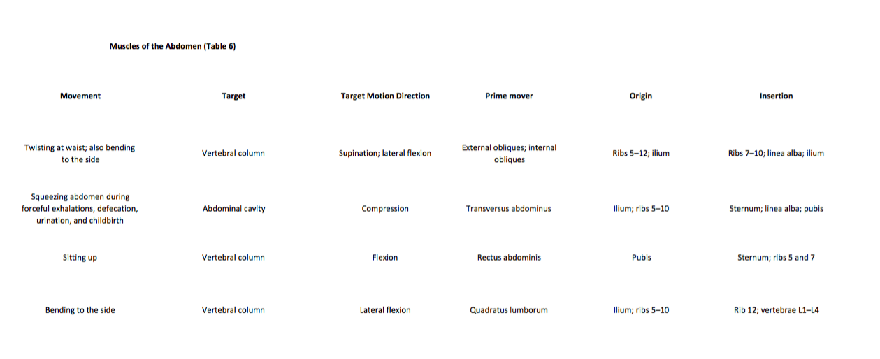
Muscles of the Abdomen
There are four pairs of abdominal muscles that cover the anterior and lateral abdominal region and meet at the anterior midline. These muscles of the anterolateral abdominal wall can be divided into four groups: the external obliques, the internal obliques, the transversus abdominis, and the rectus abdominis (Figure 1 and Table 6).

Figure 1. Muscles of the Abdomen
(a) The anterior abdominal muscles include the medially located rectus abdominis, which is covered by a sheet of connective tissue called the rectus sheath. On the flanks of the body, medial to the rectus abdominis, the abdominal wall is composed of three layers. The external oblique muscles form the superficial layer, while the internal oblique muscles form the middle layer, and the transverses abdominus form the deepest layer. (b) The muscles of the lower back move the lumbar spine but also assist in femur movements.
There are three flat skeletal muscles in the anterolateral wall of the abdomen. The external oblique, closest to the surface, extends inferiorly and medially, in the direction of sliding one’s four fingers into pants pockets. Perpendicular to it is the intermediate internal oblique, extending superiorly and medially, the direction the thumbs usually go when the other fingers are in the pants pocket. The deep muscle, the transversus abdominis, is arranged transversely around the abdomen, similar to the front of a belt on a pair of pants. This arrangement of three bands of muscles in different orientations allows various movements and rotations of the trunk. The three layers of muscle also help to protect the internal abdominal organs in an area where there is no bone.
The linea alba is a white, fibrous band that is made of the bilateral rectus sheaths that join at the anterior midline of the body. These enclose the rectus abdominis muscles (a pair of long, linear muscles, commonly called the “sit-up” muscles) that originate at the pubic crest and symphysis, and extend the length of the body’s trunk. Each muscle is segmented by three transverse bands of collagen fibers called the tendinous intersections. This results in the look of “six-pack abs,” as each segment hypertrophies on individuals at the gym who do many sit-ups.
The posterior abdominal wall is formed by the lumbar vertebrae, parts of the ilia of the hip bones, psoas major and iliacus muscles, and quadratus lumborum muscle. This part of the core plays a key role in stabilizing the rest of the body and maintaining posture.
Career Connections
Physical Therapists
Those who have a muscle or joint injury will most likely be sent to a physical therapist (PT) after seeing their regular doctor. PTs have a master’s degree or doctorate and are highly trained experts in the mechanics of body movements. Many PTs also specialize in sports injuries.
If you injured your shoulder while you were kayaking, the first thing a physical therapist would do during your first visit is to assess the functionality of the joint. The range of motion of a particular joint refers to the normal movements the joint performs. The PT will ask you to abduct and adduct, circumduct, flex, and extend the arm. The PT will note the shoulder’s degree of function, and based on the assessment of the injury, will create an appropriate physical therapy plan.
The first step in physical therapy will probably be applying a heat pack to the injured site, which acts much like a warm-up to draw blood to the area, to enhance healing. You will be instructed to do a series of exercises to continue the therapy at home, followed by icing, to decrease inflammation and swelling, which will continue for several weeks. When physical therapy is complete, the PT will do an exit exam and send a detailed report on the improved range of motion and return of normal limb function to your doctor. Gradually, as the injury heals, the shoulder will begin to function correctly. A PT works closely with patients to help them get back to their normal level of physical activity.
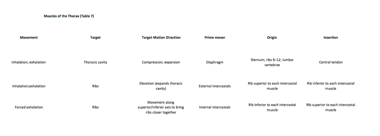
Muscles of the Thorax
The muscles of the chest serve to facilitate breathing by changing the size of the thoracic cavity (Table 7). When you inhale, your chest rises because the cavity expands. Alternately, when you exhale, your chest falls because the thoracic cavity decreases in size.
The Diaphragm
The change in volume of the thoracic cavity during breathing is due to the alternate contraction and relaxation of the diaphragm (Figure 2). It separates the thoracic and abdominal cavities and is dome-shaped at rest. The superior surface of the diaphragm is convex, creating the elevated floor of the thoracic cavity. The inferior surface is concave, creating the curved roof of the abdominal cavity.

Figure 2. Muscles of the Diaphragm
The diaphragm separates the thoracic and abdominal cavities.
Defecating, urination, and even childbirth involve cooperation between the diaphragm and abdominal muscles (this cooperation is referred to as the “Valsalva maneuver”). You hold your breath with a steady contraction of the diaphragm; this stabilizes the volume and pressure of the peritoneal cavity. When the abdominal muscles contract, the pressure cannot push the diaphragm up, so it increases pressure on the intestinal tract (defecation), urinary tract (urination), or reproductive tract (childbirth).
The inferior surface of the pericardial sac and the inferior surfaces of the pleural membranes (parietal pleura) fuse onto the central tendon of the diaphragm. To the sides of the tendon are the skeletal muscle portions of the diaphragm, which insert into the tendon while having several origins including the xiphoid process of the sternum anteriorly, the inferior six ribs and their cartilages laterally, and the lumbar vertebrae and 12th ribs posteriorly.
The diaphragm also includes three openings for the passage of structures between the thorax and the abdomen. The inferior vena cava passes through the caval opening, and the esophagus and attached nerves pass through the esophageal hiatus. The aorta, thoracic duct, and azygous vein pass through the aortic hiatus of the posterior diaphragm.
The Intercostal Muscles
There are three sets of muscles, called intercostal muscles, which span each of the intercostal spaces. The principal role of the intercostal muscles is to assist in breathing by changing the dimensions of the rib cage (Figure 3).

Figure 3. Intercostal Muscles
The external intercostals are located laterally on the sides of the body. The internal intercostals are located medially near the sternum. The innermost intercostals are located deep into both the internal and external intercostals.
The 11 pairs of superficial external intercostal muscles aid in the inspiration of air during breathing because when they contract, they raise the rib cage, which expands it. The 11 pairs of internal intercostal muscles, just under the externals, are used for expiration because they draw the ribs together to constrict the rib cage. The innermost intercostal muscles are the deepest, and they act as synergists for the action of the internal intercostals.
Muscles of the Pelvic Floor and Perineum
The pelvic floor is a muscular sheet that defines the inferior portion of the pelvic cavity. The pelvic diaphragm, spanning anteriorly to posteriorly from the pubis to the coccyx, comprises the levator ani and the ischiococcygeus. Its openings include the anal canal and urethra, and the vagina in women.
The large levator ani consists of two skeletal muscles, the pubococcygeus and the iliococcygeus (Figure 4). The levator ani is considered the most important muscle of the pelvic floor because it supports the pelvic viscera. It resists the pressure produced by contraction of the abdominal muscles so that the pressure is applied to the colon to aid in defecation and to the uterus to aid in childbirth (assisted by the ischiococcygeus, which pulls the coccyx anteriorly). This muscle also creates skeletal muscle sphincters at the urethra and anus.

Figure 4. Muscles of the Pelvic Floor
The pelvic floor muscles support the pelvic organs, resist intra-abdominal pressure, and work as sphincters for the urethra, rectum, and vagina.
The perineum is the diamond-shaped space between the pubic symphysis (anteriorly), the coccyx (posteriorly), and the ischial tuberosities (laterally), lying just inferior to the pelvic diaphragm (levator ani and coccygeus). Divided transversely into triangles, the anterior is the urogenital triangle, which includes the external genitals. The posterior is the anal triangle, which contains the anus (Figure 5). The perineum is also divided into superficial and deep layers with some of the muscles common to men and women (Figure 6). Women also have the compressor urethrae and the sphincter urethrovaginalis, which function to close the vagina. In men, there is the deep transverse perineal muscle that plays a role in ejaculation.

Figure 5. Muscles of the Perineum
The perineum muscles play roles in urination in both sexes, ejaculation in men, and vaginal contraction in women.

Figure 6. Muscles of the Perineum Common to Men and Women
REVIEW
Made of skin, fascia, and four pairs of muscles, the anterior abdominal wall protects the organs located in the abdomen and moves the vertebral column. These muscles include the rectus abdominis, which extends through the entire length of the trunk, the external oblique, the internal oblique, and the transversus abdominus. The quadratus lumborum forms the posterior abdominal wall.
The muscles of the thorax play a large role in breathing, especially the dome-shaped diaphragm. When it contracts and flattens, the volume inside the pleural cavities increases, which decreases the pressure within them. As a result, air will flow into the lungs. The external and internal intercostal muscles span the space between the ribs and help change the shape of the rib cage and the volume-pressure ratio inside the pleural cavities during inspiration and expiration.
The perineum muscles play roles in urination in both sexes, ejaculation in men, and vaginal contraction in women. The pelvic floor muscles support the pelvic organs, resist intra-abdominal pressure, and work as sphincters for the urethra, rectum, and vagina.
GLOSSARY
anal triangle
posterior triangle of the perineum that includes the anus
caval opening
opening in the diaphragm that allows the inferior vena cava to pass through; foramen for the vena cava
compressor urethrae
deep perineal muscle in women
deep transverse perineal
deep perineal muscle in men
diaphragm
skeletal muscle that separates the thoracic and abdominal cavities and is dome-shaped at rest
external intercostal
superficial intercostal muscles that raise the rib cage
external oblique
superficial abdominal muscle with fascicles that extend inferiorly and medially
iliococcygeus
the muscle that makes up the levator ani along with the pubococcygeus
innermost intercostal
the deepest intercostal muscles that draw the ribs together
intercostal muscles
muscles that span the spaces between the ribs
internal intercostal
muscles the intermediate intercostal muscles that draw the ribs together
internal oblique
flat, intermediate abdominal muscle with fascicles that run perpendicular to those of the external oblique
ischiococcygeus
muscle that assists the levator ani and pulls the coccyx anteriorly
levator ani
pelvic muscle that resists intra-abdominal pressure and supports the pelvic viscera
linea alba
white, fibrous band that runs along the midline of the trunk
pelvic diaphragm
the muscular sheet that comprises the levator ani and the ischiococcygeus
perineum
the diamond-shaped region between the pubic symphysis, coccyx, and ischial tuberosities
pubococcygeus
the muscle that makes up the levator ani along with the iliococcygeus
quadratus lumborum
posterior part of the abdominal wall that helps with posture and stabilization of the body
rectus abdominis
long, linear muscle that extends along the middle of the trunk
rectus sheaths
tissue that makes up the linea alba
sphincter urethrovaginalis
deep perineal muscle in women
tendinous intersections
three transverse bands of collagen fibers that divide the rectus abdominis into segments
transversus abdominis
deep layer of the abdomen that has fascicles arranged transversely around the abdomen
urogenital triangle
anterior triangle of the perineum that includes the external genitals
BodyFly Abs/Core Exercises
Training your abdominal muscles with constant tension is a quick way to strengthen and tone your midsection. For a great stability and core workout, try the wood chop exercise.